Additionally, we previously found an association between glaucoma pathology, which has reduced RNFL thickness, and the occurrence of dementia 3 years later ( 11). Regarding MCI and/or AD, several previous studies have demonstrated reduced RNFL thickness compared to that of a healthy control population, initially in histological studies ( 8) and then in OCT studies ( 4, 9, 10) these studies were, however, mostly cross-sectional case–control studies. This RNFL could thus enable evaluation of neurodegeneration in mild cognitive impairment (MCI) and AD pathology ( 4).Ībnormality in RNFL thickness has been described in several neurodegenerative conditions, in particular multiple sclerosis, AD, and Parkinson’s disease ( 5– 7). Spectral domain optical coherence tomography (SD-OCT) allows easy and accurate evaluation of the peripapillary retinal nerve fiber layer (RNFL), by measuring the thickness of this layer containing ganglion cell axons in a circle around the optic nerve. Thus, the condition of nerve fibers in the eye could reflect the condition of nerve fibers in the brain ( 3). The eye is a sensory organ that is truly part of the central nervous system, with neuronal cells that could be susceptible to degeneration, directly or indirectly. The retina and brain are intimately linked as a result of common embryonic origin. Detection of this neurodegenerative process at an early stage could allow prediction of cognitive decline in subsequent years. These lesions occur several years before the clinical phase of dementia ( 1, 2). RNFL thickness could reflect a higher risk of developing cognitive impairment over time.Īlzheimer’s disease (AD) is associated with brain neurodegeneration, particularly in the medial temporal area, which leads to cognitive decline and dementia. The RNFL was associated with changes in scores that assess episodic memory. No associations were found with other cognitive tests.
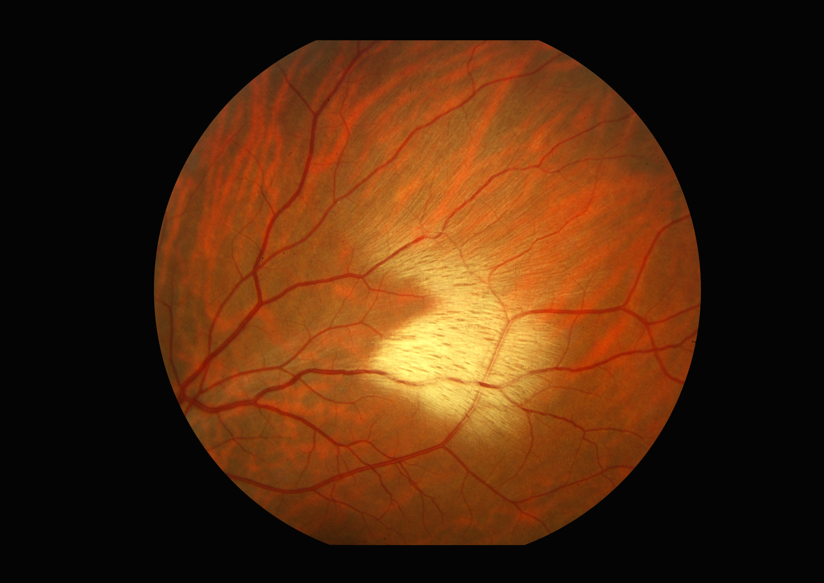
MYELINATED RETINAL NERVE FIBER LAYER FREE
Nevertheless, a thicker RNFL was significantly associated with a better cognitive evolution over time in the free delayed recall ( p = 0.0037) and free + cued delayed recall ( p = 0.0043) scores of the FCSRT, particularly in the temporal, superotemporal, and inferotemporal segments. The RNFL was not associated with initial cognitive performance. Multivariate linear mixed models were performed. RNFL was assessed at baseline by spectral domain optical coherence tomography cognitive performances were assessed at baseline and at 2 years, with the Mini–Mental State Examination, the Isaacs’ set test, and the Free and Cued Selective Reminding Test (FCSRT). Within 427 participants from the Three-City-Alienor longitudinal population-based cohort, we explored the relationship between peripapillary RNFL thicknesses and the evolution of cognitive performance. However, whether it is associated with early evolution of cognitive function is unknown. Retinal nerve fiber layer (RNFL) thickness is reduced in Alzheimer’s patients. 3University Hospital, Memory Consultation, CMRR, Bordeaux, France.2University Hospital, Ophthalmology, Bordeaux, France.1University Bordeaux, INSERM, Bordeaux Population Health Research Center, Team LEHA, UMR 1219, Bordeaux, France.

Juan Luis Méndez-Gómez 1 Marie-Bénédicte Rougier 1,2 Laury Tellouck 1,2 Jean-François Korobelnik 1,2 Cédric Schweitzer 1,2 Marie-Noëlle Delyfer 1,2 Hélène Amieva 1 Jean-François Dartigues 1,3 Cécile Delcourt 1 Catherine Helmer 1*


 0 kommentar(er)
